Have you ever noticed that when people argue about any hot topic promoted by the news media, most of them tend to fall into the trap of accepting the fundamental false premise of the topic du jour and then get tricked into debating the minutiae?
Put another way: They completely ignore the root assumptions the media is presenting and instead allow themselves to be distracted by minor details—details that tend to validate the narrative’s false framing.
Accepting a false assumption and arguing the finer points within that framework is known as “conceding the premise.”
Once it is brought to light that the underlying premise itself is unverified or unstable, the entire structural integrity of the contention collapses.
In the case of Ebola, the entire story line depends on the acceptance of a few major assumptions—with the fundamental assumption being that the “discovery” of this pathogen was legitimate in the first place and that it was this pathogen that was responsible for the deaths attributed to Ebola.
Before we tell this story, it may be helpful to readers to give a timeline of the various names of the country that was one of two places in Africa where outbreaks of Ebola were said to have occurred in 1976.
- 1885–1908: Congo Free State (established and owned by King Leopold II of Belgium)
- 1908–1960: Belgian Congo (annexed by the Belgian state)
- 1960–1964: Republic of the Congo (now an independent state; also called Congo-Leopoldville to distinguish it from the neighboring Republic of the Congo aka Congo-Brazzaville)
- 1964–1971: Democratic Republic of the Congo (the name change followed the establishment of the Luluabough Constitution)
- 1971–1997: Republic of Zaire (under dictator Mobutu Sese Seko, the country was renamed Zaire to shed colonial associations)
- 1997–Present: Democratic Republic of the Congo, sometimes shortened to DRC (this name was restored after the First Congo War and Mobutu’s removal from power)
The Discovery: Virus Hunters On Safari
In medical lore, virus hunters are Indiana Jones-like characters who track down, identify, and study “emerging pathogens” in remote and dangerous locations.
Some of the more legendary hunters of viruses are described as adventurous and eccentric daredevils who risk life and limb in “savage” lands just so they can bring home “viral trophies” in their vials.
In the case of the purported Ebola virus, its “discovery” was a collaborative effort credited to several scientists: Dr. Jean-Jacques Muyembe, Dr. Peter Piot, Dr. Guido van der Groen, and Dr. Joel Breman.
- Muyembe, a Congolese field epidemiologist, was the first to investigate an alleged Ebola outbreak in 1976 in what was at the time the Republic of Zaire. After he collected blood samples from a sick patient, he claimed to have found a new, highly lethal disease.
- Piot was at the time a young Belgian microbiologist who worked at the Institute for Tropical Medicine in Antwerp. He was credited as the first to use an electron microscope to visualize the ostensible new pathogen using Dr. Muyembe’s blood samples.
- Van der Groen and Breman, Belgian and American, respectively, were part of a team of researchers and scientists at the Institute for Tropical Medicine and the US Centers for Disease Control and Prevention (CDC). They were involved in the early lab identification and naming of the so-called virus.
Not to be omitted from this pantheon of scientific “stars” is American Dr. Karl M. Johnson, who, along with Van der Groen and Breman, is credited with naming the virus “Ebola.”
As the story goes, Ebola got its appellation when a few of the five aforementioned scientists were hanging out late one night drinking Karl’s Kentucky bourbon and discussing possible monikers for this new-to-them infection.
They presumed the virus had first surfaced in the village of Yambuku in Zaire. But, not wanting to stigmatize the villagers, Johnson suggested naming the putative pathogen after a river.
But which river? The one closest to Yambuku on the map was called “Ebola,” meaning “Black River” in the local language Lingala.
“It seemed suitably ominous,” Piot wrote. He chose the right word, “ominous”: It served as a “suitable” forewarning of what was to come, though perhaps not in the way Piot intended.
The outlandish details supplied by the scientists who said they made the momentous “discovery” of the Ebola virus in September 1976 should raise the eyebrows of any rational observer.
Their story goes something like this:
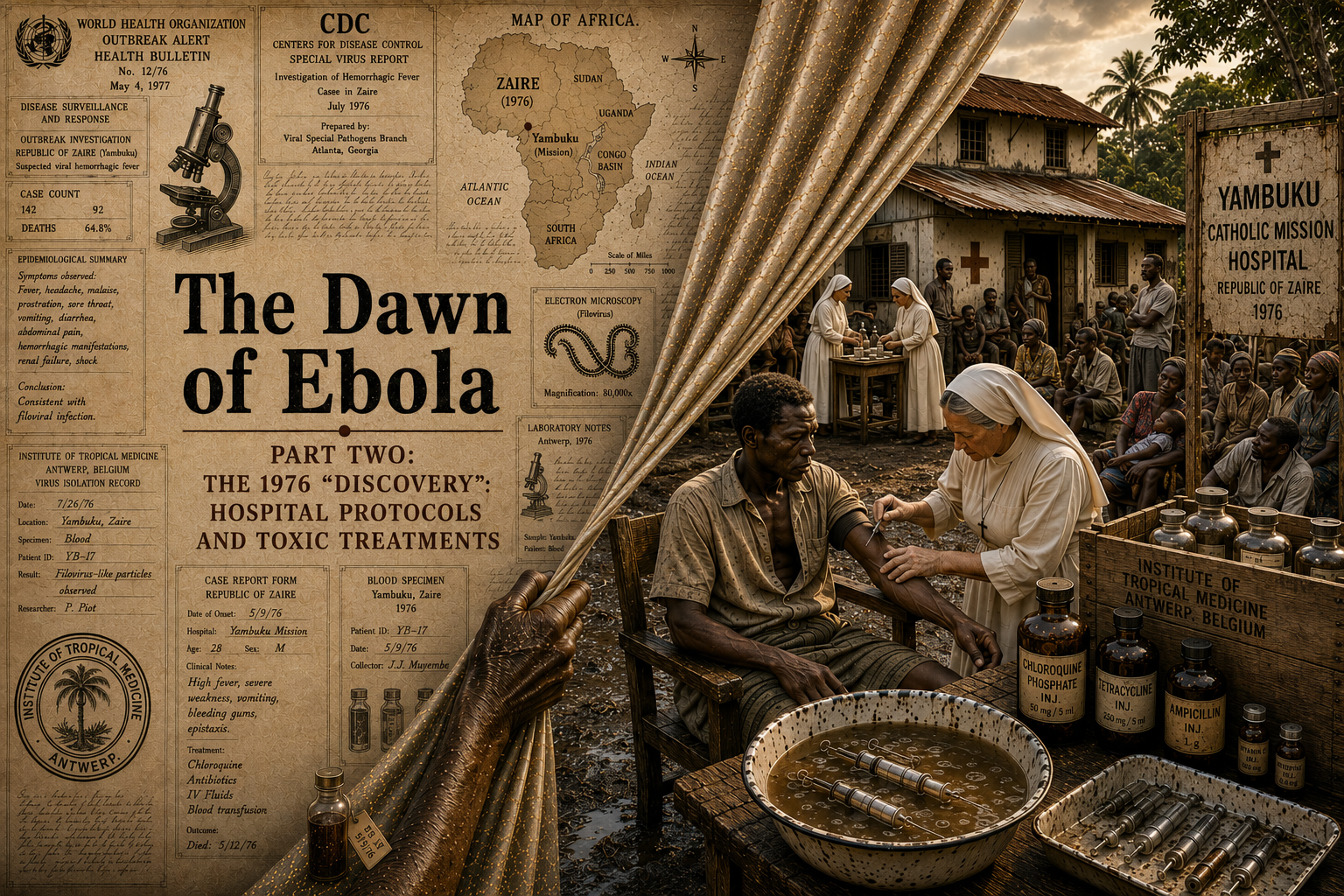
Blood was drawn from one of the dying Flemish nuns and then preserved in a Thermos flask and flown to Belgium. The sample belonged to Sister Myriam, a Belgian Catholic missionary sister stationed in Yambuku, Zaire.
A local doctor in Yambuku packaged the blood in two glass vials and placed them inside a standard glossy-blue Thermos(R) flask filled with ice cubes. The Thermos was handed to a commercial airline pilot, who flew it from Kinshasa, Zaire, directly to Antwerp, Belgium, on a passenger (not cargo) flight.
On September 29, 1976, the package was delivered to the Institute of Tropical Medicine in Antwerp (ITM), where Dr. Peter Piot and his colleagues opened it. Upon unscrewing the flask, the scientists discovered that one of the glass vials had shattered during transit. Dr. Piot et al. reached into the soupy mixture of melted ice and contaminated glass shards to fish out the single surviving, intact vial.
Unaware of the “extreme danger” and not clad in the high-containment biosafety suits used today, the Piot-led scientists team examined the specimens wearing only standard cotton lab coats and latex gloves.
Even though the ice had melted and the quality of the product had been compromised, they surprisingly claimed the “viral quality” was completely intact. Through their analysis of the remaining intact blood vial, they identified what they deemed to be spectacular, worm-like pathogens. The initial qualitative claim made by Dr. Piot on October 4, 1976—the day he first examined the sample under the electron microscope in Antwerp—was simply that the pathogen looked like a massive “worm-like” Marburg virus. But the exact numerical measurements (defining the precise differences in the length of the Marburg vs. the Ebola “worms”) were not established until more than a decade later. In 1986, the scientific community officially recognized Ebola and Marburg as distinct entities, at which point they created the Filoviridae virus family.
Despite the improper temperature and the contamination, the virus inside the blood, the Piot team said, remained “remarkably viable and deadly.” They asserted that the sample had succeeded due to its extreme viral load. Sister Myriam’s blood, they maintained, contained a staggeringly high concentration of the pathogen. They insisted this was possible because, according to them, Ebola replicates so rapidly that even a compromised, diluted sample still holds millions of “active viral particles.”
So, just to reiterate the scientists’ story, even though one glass tube shattered and created a “blood soup” at the bottom of the flask, the second glass vial miraculously remained intact. They fished this intact tube out of the slop and from this tube extracted the ostensible virus. The melted ice water inadvertently kept the sample cool enough, all the way through transport, thereby preventing the proteins from degrading completely before reaching the Institute of Tropical Medicine in Antwerp.
Strangely, it was claimed that the melted ice did not ruin “the science,” and yet it was also claimed that the biological hazard had dramatically increased when the blood mixed with the water and caused the entire inside of the Thermos flask to become a “bio-hazard splash zone.”
Please note that when Dr. Piot and his team used their hands to retrieve the intact vial from the flask, they were wearing basic latex gloves. This meant that, without realizing it, they were coating their hands with what was considered a highly concentrated, live strain of a Level 4 pathogen that would have easily soaked through the gloves and into the skin.
Miraculously, no one in the Belgian laboratory was infected or otherwise harmed by this pathogen, even though it was alleged to have a 50% mortality rate. Later, when asked about the terrifying absence of safety gear, Dr. Piot replied with a matter-of-fact “Nothing happened to any of us.” But then, later still, Piot and his teammates said they had escaped unscathed due to pure luck and to the specific transmission mechanics of “the Ebola virus.”
The Origin Story
The year was 1976. Yambuku was an isolated, impoverished village in the North Central forest of what was then the Republic of Zaire and what is now the Democratic Republic of the Congo (DRC). Not only was there no running water or electricity, but the roads connecting Yambuku to the nearest trading hub, Bumba, were mere mud tracks—poorly maintained and frequently washed away by torrential equatorial rains.
A Yambuku Catholic Mission had been established by Belgian missionaries in 1935. It was founded as part of a highly structured triple alliance between the Belgian Congo colonial state, the Roman Catholic Church, and private corporate concessions in the Belgian Congo.
The Yambuku Catholic Mission was managed by the Fathers of Scheut, a Belgian missionary order. Its Mission School was managed primarily by the Scheut priests, who employed local Congolese staff, including headmaster Mabalo Lokela. Its Mission Hospital was managed by a team of Flemish Belgian nuns. There being no resident medical doctor stationed at Yambuku in 1976, the nuns acted as the primary clinicians. They supervised a staff of some seventeen Congolese medical assistants.
Because there was no sewage system, human and animal waste contaminated Yambuku’s water sources. Villagers and missionaries routinely fell ill and died from waterborne diseases. The same untreated water supply was used to rinse the medical equipment in the hospital. No wonder rampant waterborne pathogens that caused typhoid, dysentery, and yellow fever made Yambuku’s baseline water quality highly toxic to human health.
Ebola is said to have debuted in this village around the first of September 1976. Between September 1st and October 24th, there were 318 cases of acute viral haemorrhagic fever in northern Zaire. This “outbreak” was centered in the Bumba Zone of the Equator Region, with most of the cases recorded within a 70-kilometer radius of Yambuku.
Central to the mystique of this “exotic” disease’s purported emergence is a fascinating and otherworldly explanation for its “origin.”
The WHO and the CDC ultimately settled on the bat-bushmeat theory as the leading “scientific” explanation for Ebola. According to this script, the so-called virus “spills over” from the wild into human populations via a merging of fruit bats—said to be the natural reservoir of the virus—with bushmeat (wild animal meat). The combination is said to serve as the vehicle that then carries Ebola to humans.
Though this dubious theory remains hotly contested, it is thoroughly rejected by many who consider it to be the byproduct of Western cultural myths, stereotypes, and moral judgments.
But, given the imagination of Europeans and Americans, who were conditioned to accept the emergence of an invisible threat coming from “the bush” of “deepest darkest Africa,” where “primitive things” happen, “Ebola” was an easy sell.
“Patient Zero” and Chloroquine Injections
The first recognized “case” (index case) of Ebola in Yambuku was Mabalo Lokela. On August 22, 1976, the 42-year-old headmaster of the Yambuku Mission School, a resident of Yandongi Village, was returning from a two-week driving excursion to northern Zaire and along the route allegedly purchased antelope and smoked monkey meat.
On August 26th, Lokela presented with a fever to the Yambuku Mission Hospital (YMH), where staff suspected malaria and treated him with an injection of chloroquine at its outpatient clinic. Five days later, Lokela, by then at home, grew critically ill and died on September 8. Following his funeral, 21 of Lokela’s friends and relatives fell severely ill, and 18 of them died.
Other early patients were local villagers who were visiting the outpatient clinic for routine ailments. The hospital used only five glass syringes per day. The five syringes used at the Yambuku Mission Hospital were paired with reusable metal needles, since disposable plastic syringes were not available in this remote region.
These glass syringes were reused on thousands of patients without sterilizing them between uses. The Yambuku Mission Hospital had no access to sterile water, functioning autoclaves, or disposable medical supplies. Thus, the five glass syringes allocated to the entire outpatient facility were handled through a highly dangerous routine where, after each use, the nurses placed the needle into a basin filled with warm, unsterilized surface water drawn from local streams.
At Yambuku Mission Hospital, epidemiological reports indicated that nearly all outpatients received chloroquine injections. That was the hospital’s standard procedure for any patient or staff member exhibiting a fever.
Within one week, several other persons who had received the same injections at YMH suffered what was later diagnosed as Ebola haemorrhagic fever. Almost all subsequent cases had either received injections at the hospital or had had close contact with another case. Most of these cases occurred during the first four weeks of the epidemic, after which time the hospital was closed.
A reported 11 of the 17 YMH staff members died of the disease. The documented fatality rate of the patients who received treatment at YMH was equally high. Of the 318 reported “cases,” there were 280 deaths, which represented a devastating case-fatality rate of approximately 88%.
Chloroquine is an anti-malaria drug that is known for its serious side effects. These side effects share an overlapping set of symptoms and systemic complications with what would become known as Ebola.
The primary side effects of chloroquine that present similarly to Ebola symptoms are outlined below, starting with early-stage symptoms and progressing to advanced complications:
[1] Chloroquine injections can cause nausea, vomiting, fever, and chills as side effects, which can overlap with the initial presentation of Ebola.
[2] Severe headache is another shared early symptom of chloroquine and Ebola.
[3] Chloroquine-induced muscle weakness or muscle pain also acts similarly to the body aches caused by Ebola.
[4] As the chloroquine reaction intensifies, it impacts the skin and blood systems in ways that are eerily similar to Ebola. For instance, chloroquine frequently induces severe pruritus (itching), redness, or macular rashes. Ebola has similar symptoms.
[5] Additionally, adverse reactions to chloroquine can trigger hemolytic anemia or bone marrow suppression, which presents as bruising, pale skin, or bleeding. These three symptoms mimic the early coagulopathy and bleeding abnormalities seen in Ebola.
[6] High dosage or prolonged use of chloroquine causes severe fatigue, simulating the profound prostration and lethargy seen in advanced Ebola cases.
Altogether, the symptoms for Ebola coincide fully with the side effects from a chloroquine injection. As we have noted in Part One, and as we will see repeatedly in future segments, numerous (and various) chemical toxicities will be attributed to this “new disease.”
In Yambuku Mission Hospital, almost all medication—including chloroquine for suspected malaria—was administered via parenteral injection. Parenteral injection bypasses the body’s primary protective barriers—the skin and mucous membranes—to deliver medication directly into blood vessels, muscles, or tissues. Current recommendations state that parenteral chloroquine should no longer be used because of potential toxicity.
Because chloroquine penetrates the body internally, this type of drug administration carries significant risks. Potential complications include deadly systemic blood poisoning (sepsis), life-threatening allergic reactions, shock, embolisms, nerve injury, immediate organ toxicity, cardiac arrest, and hematomas (localized bleeding).
As noted above, the unsanitary, unsafe conditions at the Yambuku Mission Hospital meant that the nursing staff administered the injections under highly compromised conditions. The five glass syringes and metal needles used each day to treat hundreds of patients were not only unsterilized, they were so badly, so barely cleansed that they often held the last patient’s blood on the tip when given to the next patient.
The significance of what was happening at this mission hospital and others cannot be overstated. In fact, the majority of the aforementioned illnesses seemed to have originated inside the hospitals.
Granted, the official World Health Organization (WHO) International Commission report does not reveal the total number of hospitalized patients who died. But historical data shows that virtually every single one of the 280 deaths had a direct link to the hospital. Roughly 85 to 100 of the deceased can be directly tracked back to being injected or treated inside the Yambuku Hospital Mission facility itself.
Dumping Toxic Beta-Lactam Antibiotics in Africa
During the 1970s and 1980s, there was intense international controversy surrounding Western pharmaceutical companies dumping banned or substandard drugs in African, Asian, and Latin American countries—drugs that were unapproved, or at least heavily restricted, in their home countries.
A classic example was chloramphenicol, which is a non-beta-lactam antibiotic. Chloramphenicol was severely restricted or banned for routine use in Europe and the US throughout those two decades because it could cause fatal aplastic anemia. Yet it was aggressively marketed and widely distributed over the counter in many African nations.
Broad-spectrum beta-lactams like ampicillin and older cephalosporins were frequently sent to African nations as “humanitarian aid” or sold cheaply by brokers after these drugs had expired or failed quality control checks (often the result of contamination during formulation) in European facilities.
European regulatory bodies fiercely policed these products and prevented the rejects from being used domestically. But they allowed the same products to be manufactured for export if the receiving country’s laws did not explicitly forbid them. Rather than destroy the failed batches and take a financial hit, pharmaceutical companies sold these “off-spec” lots to corrupt international brokers at steep discounts. Then the brokers marketed them to under-regulated ministries of health or to private wholesalers in Africa.
A significant portion of failed or expiring European beta-lactams crossed national borders disguised as “humanitarian aid.” European pharma firms discovered they could claim substantial domestic tax deductions by donating excess inventory to developing nations. European warehouses would clear near-expired or poorly stored ampicillin and older cephalosporins and ship them to Africa in bulk barrels. The shipments often lacked proper storage instructions and labels translated into the right language and even labels with the correct product names on them. Being highly vulnerable to environmental factors, these broad-spectrum beta-lactams, which had already failed quality checks in a temperate European facility, would deteriorate further when shipped to sub-Saharan Africa.
A landmark 1982 study published in PubMed detailed how multinational pharma firms systematically targeted Zaire during this time frame. It pointed out that major European firms used Zaire as a market to dump highly problematic drugs that were restricted or rarely used in Western industrialized countries. And it noted that missionaries and local health workers routinely administered these volatile, fast-degrading broad-spectrum antibiotics to the public.
More specifically, Belgian Catholic missionary networks and humanitarian entities heavily utilized these substandard or expired pharmaceuticals in their hospitals across Zaire during the 1970s and 1980s. Belgian nuns and doctors routinely received massive crates of broad-spectrum antibiotics—primarily ampicillin, amoxicillin, and early cephalosporins—that were either near the expiration date, completely expired, or rejected from European clinical distribution due to production non-compliance.
All of these shenanigans were couched in the language of “humanitarian assistance.”
The administration of substandard and expired beta-lactams to Africans during this era had catastrophic health consequences. When beta-lactam antibiotics (especially penicillins) degrade due to heat, humidity, or poor manufacturing quality control, they break down into dangerous chemical sub-products. Thus:
- The degradation products of ampicillin triggered severe, sometimes fatal allergic reactions (anaphylaxis).
- Contaminated batches of these antibiotics could contain chemical impurities that would have caused acute kidney injury or severe bone marrow suppression.
- Patients who were given the injectable form of degraded beta-lactams with unsterile equipment regularly experienced painful bacterial abscesses at the injection site.
- In severe cases, patients suffered tissue necrosis (tissue death) and gangrene, requiring limb amputations in rural clinics that lacked proper surgical facilities.
Toxic accumulation, or “poisoning,” with beta-lactam antibiotics is known to lead to several specific medical conditions characterized by moderate-to-life-threatening hemorrhaging. Under the best of circumstances, known side effects include spontaneous internal or external hemorrhages, large hematomas, severe epistaxis (nosebleeds), life-threatening gastrointestinal or intracranial hemorrhages, massive amounts of bloody diarrhea, severe abdominal cramping accompanied by severe intestinal bleeding, and other bleeding disorders.
It is well known that these bleeding conditions can absolutely occur as a result of using banned, unregulated, or counterfeit beta-lactams—and, in many cases, the danger is often significantly higher. Banned or black-market antibiotics that were routinely smuggled across borders in hot, humid, and unrefrigerated shipping containers contained a high concentration of degraded toxins. Beta-lactam rings are chemically unstable and break down quickly if not stored with precise climate controls.
By the time the compromised drug reached patients in Zaire, it had degraded into a cocktail of highly reactive chemical sub-products. These foreign degradation compounds were apt to violently trigger drug-induced immune thrombocytopenia (DITP), leading to spontaneous hemorrhages.
These banned and highly degraded beta-lactams fostered bleeding conditions that occurred through several pathways. Counterfeit or poorly manufactured batches frequently suffered from “bad mixing” during production. One pill might contain almost no active ingredient, while the next pill or vial contained a massive, toxic overdose triggering rapid internal bleeding. A patient given a substandard beta-lactam contaminated with toxic solvents was likely to also experience sudden acute kidney injury.
These beta-lactam antibiotics, most notably injectable penicillin, were routinely used by the medical team at the Yambuku Mission Hospital in 1976. They were a staple of the remote hospital’s daily operations and were used for a wide variety of bacterial infections, wounds, and systemic febrile illnesses.
Importantly, all these symptoms mirror the tragic mass hemorrhages said to be a feature of “Ebola” in its advanced stages.
Given what we know about European nations dumping these dangerous beta-lactams in Africa, specifically targeting Zaire and known to be used regularly by Belgian missionaries in their remote mission hospitals in Zaire, is it not logical to conjecture that there had to have been some disastrous consequences for the locals who were subjected to these toxic injections?
And isn’t it also quite logical to theorize that, since manifestations of “Ebola” and symptoms of beta-lactam poisoning run parallel to one another, there might have been more at work than merely an invisible microbe in the disease named “Ebola” in Yambuku?
Toxic Burials and Deadly Fumigation
A 1978 WHO Bulletin chronicles how phenol and formaldehyde became twin pillars of the environmental decontamination strategy to eliminate “Ebola.”
Traditional burials in Yambuku were deeply spiritual, community-driven events that required hands-on handling of the deceased. Local customs dictated that a proper farewell required physical touch to honor the dead and ensure their spirit transitioned peacefully to the ancestral world.
As a consequence of the supposed outbreak of “Ebola,” international health teams completely banned traditional burials and replaced them with clinical, chemical-based interment protocols.
Under these untraditional protocols, the bodies of the victims were wrapped before burial in cotton sheets that were heavily impregnated with a phenolic disinfectant. Responders sealed the rooms of deceased patients and generated formaldehyde gas on-site (often by heating paraformaldehyde or mixing formalin with potassium permanganate). They repeated this intensive fumigation process over four consecutive days.
You can imagine that fumigating sealed rooms with formaldehyde gas for four consecutive days and wrapping deceased bodies in phenol-impregnated cotton sheets would be an extraordinary level of chemical exposure for medical workers, family members, and returning villagers. Both of these chemicals are highly toxic, corrosive agents.
Formaldehyde gas is a severe respiratory and tissue irritant. Heavy exposure inside unventilated homes causes severe respiratory distress, intense burning of the eyes, chemical burns to the lining of the nose, splitting headaches, and nausea from inhaling the fumes.
Phenolic disinfectants, for their part, were heavily formulated in the 1970s with aggressive, first-generation compounds like pure phenol (carbolic acid) and cresols (coal tar derivatives). Because these formulas of fifty years ago were far more crude and concentrated than modern versions, they carried severe health risks.
These 1970s phenols were highly fat-soluble and passed rapidly through intact skin directly into the bloodstream. Once absorbed, the chemical caused “carbol marasmus” and systemic poisoning. This led to central nervous system depression, sudden drop in blood pressure (cardiovascular collapse), severe kidney damage, and dark green or black urine. Moreover, inhaling heavy phenol vapors in poorly ventilated spaces caused acute neurological symptoms.
Such a massive deployment of chemicals added yet another layer of toxic exposures faced by Yambuku villagers and medical staff.
Families who followed instructions to disinfect and temporarily leave their homes returned to closed houses trapped with heavy, toxic vapors. Anyone who moved back into a heavily fumigated house or who handled a body soaked in phenol quickly developed severe respiratory distress, vomiting, skin lesions, and physical collapse. An immediate collapse from respiratory failure and vomiting reinforced the belief that the hospital’s medicine—and the containment process itself—was a death sentence. Naturally, this belief caused many residents to flee their village.
Of course the medical industry ignored—and continues to ignore—the obvious fact that the clinical manifestations of acute phenol exposure almost perfectly matched what would retroactively and questionably be categorized as the advanced stages of “Ebola haemorrhagic fever.” The many horrible symptoms were—and still are—explained away as the “inadvertent spreading of the virus.” But is it possible that these medical “experts” have been concealing or ignoring the devastating effects of their own medical interventions on innocent civilians? Such deceit, if deliberate, is unethical. Such harm, if intentional, is unconscionable.
Two Reports and More Questions
In 1977, a three-day colloquium between the WHO and a large international contingent of members of the medical and science community was held in Belgium. Together, they produced a 280-page report, “Ebola virus haemorrhagic fever: proceedings of an International Colloquium on Ebola Virus Infection and other Haemorrhagic Fevers.”
A careful reader of this report will find plenty of clues that will in turn elicit some hard questions about what actually happened in Zaire (and elsewhere in Africa) in 1976—particularly in the hospitals.
For example, the report noted on page 93 that in the village of Yamolembia, located five kilometers from Yambuku, between September 4 and October 18, 1976, “24 persons developed probable or confirmed cases of EbHF.” Of particular interest is that “[t]he first case was a 27 year old man who received an injection at YMH outpatient clinic on 29 August,” and that “[w]ithin six days, four more persons with a history of injection at the Yambuku Mission Hospital became ill.” [Emphasis added.]
On page 94, the report observed:
“People in the community had already associated the mission hospital with the epidemic and had stopped coming to the outpatient department.”
On page 95, the report speculated:
“The source of this epidemic, although not determined exactly, probably originated in Sudan,” and “The origin of the cases in Sudan is still obscure. This epidemic was also associated with a hospital.” [Emphasis added.]
An official bulletin released in 1978 by the WHO reported that in early September 1976, several dozen patients who had received injections at YMH developed a similar febrile hemorrhagic syndrome and died in about one week. As panic set in, patients fled the hospital to avoid the “mystery disease.”
Although official historical records do not track the exact number of patients who died while physically inside the hospital building, we know that the vast majority of the region’s “Ebola” deaths had a direct connection to that mission hospital. And, as observed on page 116 of the 1977 report from the international colloquium, the “transmission” of Ebola was “interrupted by the closure of Yambuku Hospital with cessation of giving injections.” [Emphasis added.] In other words, once the hospital was closed down, the “epidemic” disappeared with it.
The Other 1976 “Outbreak”
In the late summer of 1976, another mysterious “outbreak” of haemorrhagic fever (subsequently named “Ebola haemorrhagic fever”) occurred in southern Sudan. [NOTE: As of July 9, 2011, the southern region of Sudan became the independent Republic of South Sudan.] The outbreak in the Sudanese “source town” of Nzara was said to have originated with workers at a cotton factory. The disease supposedly spread to the Sudanese village of Maridi, some 95 miles away, where it reportedly was amplified in a large, active hospital.
Because the symptoms—including high fever, abdominal pain, and severe toxemia—closely mirrored those of a severe bacterial infection, health officials and medical teams initially diagnosed the epidemic as typhoid fever. This prompted local health authorities to enact several emergency measures to contain what they believed to be a massive typhoid outbreak. One such measure was the mass vaccination campaign against typhoid they initiated in September. [Emphasis added.]
During this gigantic emergency campaign, 13,914 doses of typhoid vaccine (as well as cholera vaccines) were administered as a countermeasure.
Coinciding with this mass vaccination campaign in southern Sudan, the mortality rate of hospitalized patients increased from 25% in August to 44.6% in September and to 70% in October. The October spike came after the massive vaccination campaign was launched in September.
On the same page, D.P. Francis also observed:
“There were people infected in the hospitals, admitted initially for various diseases, who were treated in the hospitals, including injections and who 5 to 7 days later came down with usually fatal disease.”
The report omitted the active ingredients of those routine hospital injections.
Again on page 115, another co-author, M. Isaäcson, commented:
“The Maridi Hospital was a large teaching hospital and it was noted that the main victims were the student nurses.”
On page 116, the authors wrote:
“A typhoid vaccination campaign was prescribed for all medical personnel [meaning nurses] and the population at risk.”
As it turned out, 33 of the 61 nurses at the Maridi Hospital died, which begs the question: Could a typhoid vaccination campaign also have been part and parcel of the deadly hospital protocols in Yambuku, Zaire?
Unlike in Maridi, where the hospital continued operating long enough to coordinate a late-September vaccine drive, the Yambuku Mission Hospital had completely collapsed before mid-September. By then, as you will remember, 11 of the Yambuku Mission Hospital’s 17 medical staff members had already died. There was no functioning medical infrastructure left at YMH to organize or administer a vaccine campaign.
Yet, it is highly likely that individuals in Yambuku did receive typhoid vaccines earlier in 1976 as part of routine, ongoing medical care, as observed in archival and epidemiological reports that show typhoid vaccination was actively happening in the area earlier that year.
The issues surrounding the deployment of typhoid vaccines are of particular concern. We say that because the specific type of typhoid vaccine administered to the villagers, missionaries, and health staff in Yambuku in 1976 was the inactivated whole-cell typhoid vaccine. It was often combined as the “TAB” vaccine for typhoid and paratyphoid A and B.
This vaccine was eventually discontinued and phased out globally due to its severe systemic side effects. The CDC acknowledged those side effects when it formally recommended the discontinuation and removal of this typhoid vaccine from the market in 1994.
Although this history has largely “disappeared” from the annals of the “acceptable versions” of the Ebola story, as told by the Western world, it has not been lost to the African locals, who have vowed to stay away from the deadly hospitals and the toxic injections during the current Ebola scare.
This is Part Two of our Ebola Series. Read Part One | Part Three. Stay tuned for Part Four.